


 HOME
HOME
In the past, MRI was not very reliable for diagnosing cerebral hemorrhage in the acute phase (within 3 days), and CT was the main method of diagnosis. Recently, a diagnostic imaging method for MRI called T2*-weighted imaging (also called T-to-star-weighted imaging) has been developed, and intracerebral hemorrhage can be clearly visualized by MRI in the acute phase (Fig. 1). ,Figure 2).
On the contrary, the T2*-weighted image clearly shows the old cerebral hemorrhage that could not be seen with CT. Even in patients who have had a cerebral hemorrhage for the first time, a small cerebral hemorrhage can be found in a considerable percentage of cases when T2*-weighted images are taken.
In other words, if T2*-weighted imaging is performed during a brain checkup, cerebral hemorrhage can be detected before symptoms develop and large cerebral hemorrhage can be prevented in the future.
Immediately after this image was introduced, I compared head CT and T2*-weighted images of patients with cerebral hemorrhage, analyzed their characteristics, and made a conference report and paper presentation.
This page contains a part of the contents of the paper presentation.
I think this is an easy-to-understand example of how the development of new imaging methods can have a major impact on the diagnosis and treatment of stroke.
CIReserch31:21-27, 2009(JAPANESE)
 CI Reserch 2009
Clinical Neuroscience 28:3, p346-347, 2010(JAPANESE)
 Clinical Neuroscience 2010
Multiple hypointense lesions on MRI T2*-weighted images of intracerebral hemorrhage
Features of T2*-weighted imaging: T2*-weighted imaging is a type of MRI imaging technique that is sensitive to magnetic field inhomogeneities, using the gradient echo technique. T2* is strongly affected by the magnetic susceptibility effect of the local magnetic field, and is markedly shortened by bleeding, the presence of a contrast agent, etc. If the magnetic susceptibility effect is too sensitive, it will be affected by various artifacts (signal distortion), making it difficult to distinguish it from acute intracerebral hematoma.
Advantages of T2*-weighted images: New and old hemorrhagic lesions can be visualized well, and the ability to detect microhemorrhagic foci in microhemorrhages, hemangioma, brain surface hemosiderosis, and diffuse axonal injury is significantly superior to other images.The imaging time is short, and it has the advantage of being easy to perform even for patients with body movements and restlessness. In particular, the diagnosis of occult intracerebral hemorrhage has become easier than before (Fig. 2). In patients with amyloid angiopathy and hypertension, old hemorrhages that are not clear on CT or conventional MRI may be clearly visualized only on T2*-weighted images, and in stroke, the latent hemorrhage is more latent than previously thought. Recent reports have revealed that there are many cases with sexual microbleeds. Occult intracerebral hemorrhage: T2*-weighted images are excellent in locating occult intracerebral hemorrhage (Fig. 2). We examined T2*-weighted images of patients with intracerebral hemorrhage and reported that 92% had microbleeds antecedent to other sites. Kato et al. examined T2*-weighted images and found that microintracerebral hemorrhage was observed in 71.4% of patients with intracerebral hemorrhage, 62.1% of lacunar infarction, 30.4% of cardiogenic cerebral embolism, 20.8% of atherothrombotic cerebral infarction, and controls. But 7.7% reported seeing it. It has been reported that when multiple occult microhemorrhages are seen on T2*-weighted images, higher functional disorders are likely to be seen from a young age. In addition, it has been found that many cases of symptomatic intracerebral hemorrhage occur during repeated microhemorrhages. During outpatient examinations and brain checkups, in addition to conventional MRI imaging methods such as T1-weighted images, T2-weighted images, and FLAIR images, T2*-weighted images were used to diagnose the presence of occult intracerebral hemorrhage, and occult intracerebral hemorrhage was confirmed. Patients are recommended to take preventive measures against lifestyle-related diseases, including strict control of blood pressure. Disadvantages of T2*-weighted images: Compared to conventional imaging methods, T2*-weighted images emphasize low signal (signal loss) such as air, calcifications, skulls, flow voids, and metals, and it is important to distinguish them from these. (Fig. 3).
Conclusion: T2*-weighted imaging has made it possible to diagnose intracerebral hemorrhage in the acute phase using MRI, which was previously difficult, and it has also become possible to detect minute intracerebral hemorrhage in the chronic phase. However, it is necessary to pay attention to the drawbacks of T2*-weighted images (susceptibility to magnetic susceptibility artifacts, inability to accurately grasp the shape of the hematoma due to the existence of areas with different signal intensities within the hematoma, etc.).
Figure 1
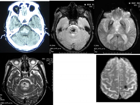
Head CT, MRI T2*-weighted image, T2-weighted image of acute pontine hemorrhage.
In the T2*-weighted images (three images on the right), the acute hemorrhage appears black around and white inside, while the hidden cerebral hemorrhage looks like a ball of black thread.
Figure 2
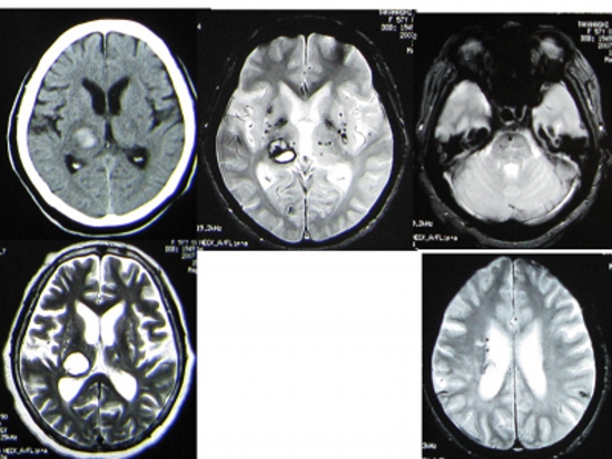
Head CT, MRI T2*-weighted image, T2-weighted image of right thalamic hemorrhage.
T2*-weighted images (right 3 images) show that there were already more than 20 small hidden cerebral hemorrhages before this hemorrhage.
Figure 3
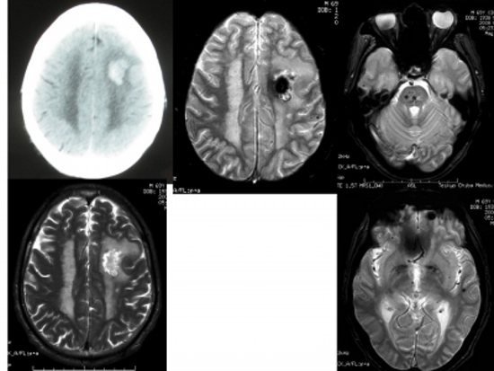
Head CT, MRI T2*-weighted image, T2-weighted image of left frontal subcortical hemorrhage.
T2*-weighted images (three images on the right) show numerous hidden cerebral hemorrhages in the brain stem, left frontal lobe, and left temporal lobe.
Figure 4
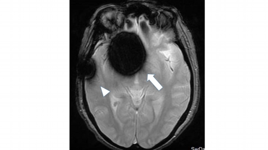
Shows artifacts (signal disturbances) that are a drawback of T2*-weighted images.
After clipping an aneurysm aneurysm, there is a huge spherical low-signal area around the aneurysm clip (arrow), and the titanium cranioplasty plate on the right temporal region exhibits metal artifacts, making the surrounding lesions undiagnosable. (arrowhead).
 HOME JAPANESE
 Self Introduction
 Paper/Presentation